Brain
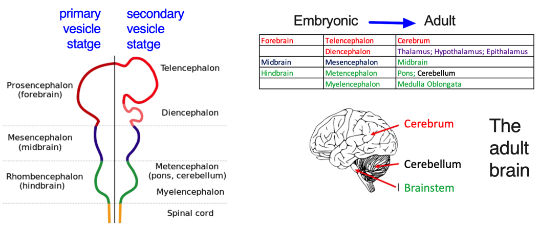
The embryonic brain develops through the enlargement of neural tube vesicles, first forming three primary vesicles and later five secondary vesicles.
For further details, you can visit this link.

In general, the adult brain can be categorized into four main parts: the cerebrum, brainstem, cerebellum, and diencephalon.
- The cerebrum develops directly from the telencephalon.
- The cerebellum forms from the metencephalon, along with the pons.
- The brainstem consists of the midbrain, pons, and medulla oblongata.
- The diencephalon gives rise to important structures such as the thalamus, hypothalamus, and epithalamus.
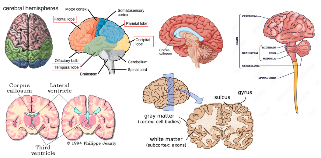
Functional areas of cerebral cortex (Images taken from Budday S et al., Sci Rep, 2014; Brodal, P. The central nervous system. Oxford University Press).

- The cerebrum consists of two cerebral hemispheres separated by the longitudinal fissure.
- The corpus callosum forms the floor of the longitudinal fissure, separating the two cerebral hemispheres.
- Part of the corpus callosum also forms the roof of the lateral ventricles.
- The lateral ventricles are the two largest ventricles in the brain and contain cerebrospinal fluid (CSF).
A sulcus is a depression or groove in the cerebral cortex while a gyrus is a ridge-like elevation found on the surface of the cerebral cortex.
Gray matter (GM) consists of neuronal cell bodies and is responsible for processing information, while white matter (WM) comprises myelinated axons that connect different brain regions. Changes in GM and WM volumes are significant in diagnosing neurological conditions. For instance, WM loss occurs in multiple sclerosis due to axonal damage, and GM volume decreases in neurodegenerative diseases like dementia and amyotrophic lateral sclerosis (ALS), where motor neurons die, especially in the spinal cord.
The two primary categories of cells in the brain are neurons and glial cells.

A “brain cell” is a broad term that often specifically refers to neurons, while “nuclei” refers to clusters of cell bodies in the CNS, which are organized based on functional similarities.
These nuclei can be broadly categorized into two types:
Subcortical nuclei: Located deep within the brain, beneath the cerebral cortex. Examples include the basal ganglia (involved in motor control), the thalamus (a sensory relay center), and the hypothalamus (which regulates vital functions like hunger and sleep).
Cortical nuclei: Found in the cerebral cortex, the brain’s outer layer. These include sensory nuclei (which process input from the senses), motor nuclei (which control movement), and association nuclei (which integrate different types of information).
Further Reading:
1.Basal Ganglia Motor Circuit
2.Basal Ganglia Circuitry in Psychiatric Disorders
3.Basal Ganglia Disorders
4.What Are Striosomal and Matrix Compartments
