Basal Ganglia Circuits
The basal ganglia is a network of subcortical brain structures distributed across the telencephalon, diencephalon, and mesencephalon (midbrain).
The basal ganglia were once believed to be involved only in motor control, but we now know that dysfunction in its various regions and circuits contributes to a range of clinical issues beyond just movement disorders.
Basal ganglia circuits can be broadly divided into three functional loops: the motor/sensorimotor, associative/cognitive, and limbic circuits.  DLS, dorsolateral striatum; DMS, dorsomedial striatum; GPi, globus pallidus internal section; MD, medial dorsal thalamus; NAc, nucleus accumbens; SNc, substantia nigra pars compacta; SNr, substantia nigra pars reticulata; VA, ventral anterior thalamus; VL, ventrolateral thalamus; VP, ventral palidum; VTA, ventral tegmental area,
By forming circuits with the cerebral cortex and thalamus, the basal ganglia help regulate voluntary movements, learning, decision-making, and motivation. Key components include
- Striatum:
- Caudate nucleus
- Putamen
- Ventral striatum (includes the nucleus accumbens, involved in reward processing)
- Globus Pallidus:
- Internal segment (GPi)
- External segment (GPe)
- Ventral pallidum (involved in reward and motivation)
Subthalamic nucleus: Crucial for regulating movements and motor output.
- Substantia nigra:
- Pars compacta (SNc): Produces dopamine, essential for motor control.
- Pars reticulata (SNr): Sends inhibitory signals, mainly to the thalamus.
The striatum is the largest component of the basal ganglia and acts as the primary hub for receiving inputs (afferent projections) from multiple regions of the brain including from cortex, thalamus, hippocampus, and brainstem. The striatum has two main compartments: striosomes (or patches) and the matrix. Striosomes are small clusters rich in dopamine D2 receptors, substance P, and enkephalin, linking them to the limbic system and roles in emotion and reward. In contrast, the matrix, which surrounds the striosomes, contains high levels of calbindin, somatostatin, and acetylcholinesterase, connecting it more to the sensory-motor system and movement coordination. Together, these compartments allow the striatum to integrate emotional and motor functions.
- Key Afferent Inputs to the Striatum:
- Cerebral Cortex: The cortico-striatal pathway sends sensory and motor information from the cortex to the striatum, where it is processed to initiate and coordinate voluntary movements.
- Thalamus: The thalamostriatal pathway delivers modulatory signals to the striatum, impacting cognitive functions and motor coordination.
- Substantia Nigra Pars Compacta (SNc): Dopaminergic neurons from the SNc project to the striatum. This nigrostriatal pathway modulates the striatum’s activity, particularly affecting reward processing and fine-tuning motor control.
- Primary Efferent Outputs from the Striatum:
- Globus Pallidus (GP): The striatum sends output signals to both the internal and external segments of the globus pallidus, which are critical for controlling the motor output pathway.
- Substantia Nigra Pars Reticulata (SNr): The striatum also projects inhibitory signals to the SNr, which helps regulate motor function by influencing the thalamus.
These interconnected pathways allow the striatum to act as a central relay station for regulating movement, behavior, and cognitive processes. It processes information from various brain regions and sends feedback to ensure smooth and coordinated motor functions.
Basal ganglia: Direct and indirect pathways (motor circut)
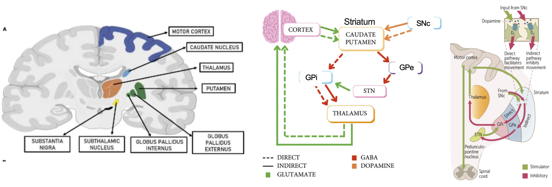
(Images taken from Rocha GS et al., Front Syst Neurosci. 2023; Audi A et al., Case Rep Neurol Med. 2023).  SNc, substantia nigra pars compacta; SNr, subtantia nigra pars reticulata; GPe, globus pallidus externus; GPi, globus pallidus internus; STN, subthalamic nucleus
The striatum contains a main type of neuron called the GABAergic spiny projection neuron (SPN), also known as the medium spiny neuron (MSN). These neurons are key to the basal ganglia and are divided into two groups: direct pathway neurons (dSPNs) and indirect pathway neurons (iSPNs). About 90% of the neurons in the striatum are MSNs that express either dopamine receptor D1 or D2, and they have opposite roles in movement control. D1-MSNs, part of the direct pathway, help promote movement by sending signals to the globus pallidus interna (GPi) and substantia nigra reticulata (SNr). In contrast, D2-MSNs, which are part of the indirect pathway, work to reduce movement by projecting to the globus pallidus externa (GPe).
Direct Pathway: The direct pathway begins in the cortex, where excitatory signals are sent to the striatum (caudate and putamen). These structures then send inhibitory signals to the globus pallidus internus (GPi), which inhibits the thalamus. Since the thalamus is responsible for movement, inhibiting an inhibitor results in overall stimulation of movement. Dopamine from the substantia nigra acts on this pathway via D1 receptors, further promoting movement.
Indirect Pathway: The indirect pathway also starts with excitatory signals from the cortex to the striatum (caudate and putamen). In response, the striatum send inhibitory signals to the globus pallidus externus (GPe), which normally inhibits the subthalamic nucleus (STN). When the GPe is inhibited, the STN becomes more active and stimulates the globus pallidus internus (GPi). This leads to the GPi inhibiting the thalamus, resulting in suppression of movement. Dopamine from the substantia nigra influences this pathway through D2 receptors, reducing its inhibitory effects on movement
Note: The SNr (substantia nigra pars reticulata) and GPi (globus pallidus internus) are both crucial output centers in the basal ganglia. However, the GPi is typically emphasized for controlling movements of the body, such as the limbs and trunk, while the SNr plays a bigger role in eye movements and head posture. This distinction is why the SNr is sometimes left out of certain models focused on general body movement.
Further Reading:
1.Basal Ganglia Motor Circuit
2.Basal Ganglia Circuitry in Psychiatric Disorders
3.Basal Ganglia Disorders
4.What Are Striosomal and Matrix Compartments
