
Brain & Pathways of Movement
The embryonic brain develops through the enlargement of neural tube vesicles, first forming three primary vesicles and later five secondary vesicles.
For further details, you can visit this link.
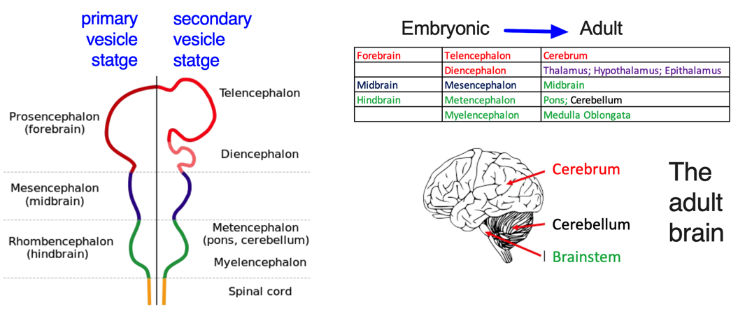
In general, the adult brain can be categorized into four main parts: the cerebrum, brainstem, cerebellum, and diencephalon.
- The cerebrum develops directly from the telencephalon.
- The cerebellum forms from the metencephalon, along with the pons.
- The brainstem consists of the midbrain, pons, and medulla oblongata.
- The diencephalon gives rise to important structures such as the thalamus, hypothalamus, and epithalamus.
Functional areas of cerebral cortex (Images taken from Budday S et al., Sci Rep, 2014; Brodal, P. The central nervous system. Oxford University Press).
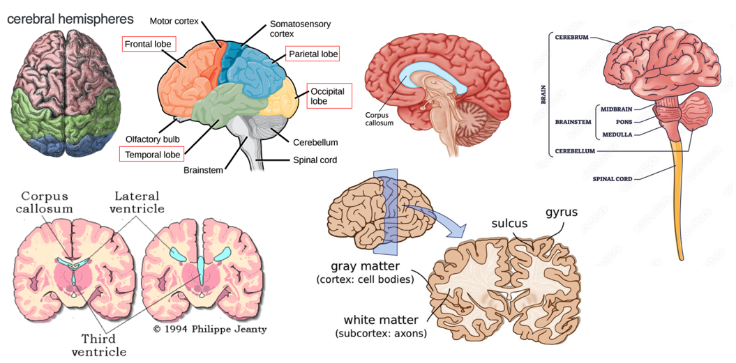
- The cerebrum consists of two cerebral hemispheres separated by the longitudinal fissure.
- The corpus callosum forms the floor of the longitudinal fissure, separating the two cerebral hemispheres.
- Part of the corpus callosum also forms the roof of the lateral ventricles.
- The lateral ventricles are the two largest ventricles in the brain and contain cerebrospinal fluid (CSF).
A sulcus is a depression or groove in the cerebral cortex while a gyrus is a ridge-like elevation found on the surface of the cerebral cortex.
Gray matter (GM) consists of neuronal cell bodies and is responsible for processing information, while white matter (WM) comprises myelinated axons that connect different brain regions. Changes in GM and WM volumes are significant in diagnosing neurological conditions. For instance, WM loss occurs in multiple sclerosis due to axonal damage, and GM volume decreases in neurodegenerative diseases like dementia and amyotrophic lateral sclerosis (ALS), where motor neurons die, especially in the spinal cord.
The two primary categories of cells in the brain are neurons and glial cells.
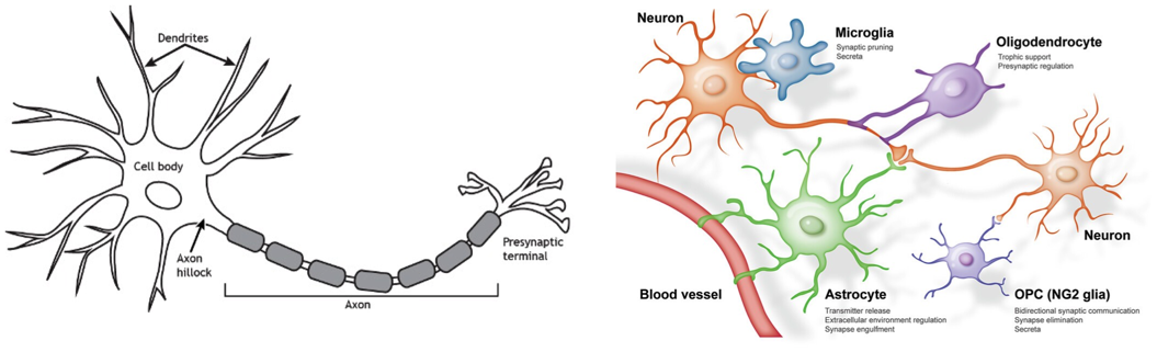
A “brain cell” is a broad term that often specifically refers to neurons, while “nuclei” refers to clusters of cell bodies in the CNS, which are organized based on functional similarities.
These nuclei can be broadly categorized into two types:
Subcortical nuclei: Located deep within the brain, beneath the cerebral cortex. Examples include the basal ganglia (involved in motor control), the thalamus (a sensory relay center), and the hypothalamus (which regulates vital functions like hunger and sleep).
Cortical nuclei: Found in the cerebral cortex, the brain’s outer layer. These include sensory nuclei (which process input from the senses), motor nuclei (which control movement), and association nuclei (which integrate different types of information).
The basal ganglia is a network of subcortical brain structures distributed across the telencephalon, diencephalon, and mesencephalon (midbrain). By forming circuits with the cerebral cortex and thalamus, the basal ganglia help regulate voluntary movements, learning, decision-making, and motivation. Key components include
- Striatum:
- Caudate nucleus
- Putamen
- Ventral striatum (includes the nucleus accumbens, involved in reward processing)
- Globus Pallidus:
- Internal segment (GPi)
- External segment (GPe)
- Ventral pallidum (involved in reward and motivation)
Subthalamic nucleus: Crucial for regulating movements and motor output.
- Substantia nigra:
- Pars compacta (SNc): Produces dopamine, essential for motor control.
- Pars reticulata (SNr): Sends inhibitory signals, mainly to the thalamus.
The striatum is the largest component of the basal ganglia and acts as the primary hub for receiving inputs (afferent projections) from multiple regions of the brain. It is closely connected with the cerebral cortex, limbic system, and thalamus, making it essential for regulating motor control, cognition, and emotional behaviors.
- Key Afferent Inputs to the Striatum:
- Cerebral Cortex: The cortico-striatal pathway sends sensory and motor information from the cortex to the striatum, where it is processed to initiate and coordinate voluntary movements.
- Thalamus: The thalamostriatal pathway delivers modulatory signals to the striatum, impacting cognitive functions and motor coordination.
- Substantia Nigra Pars Compacta (SNc): Dopaminergic neurons from the SNc project to the striatum. This nigrostriatal pathway modulates the striatum’s activity, particularly affecting reward processing and fine-tuning motor control.
- Primary Efferent Outputs from the Striatum:
- Globus Pallidus (GP): The striatum sends output signals to both the internal and external segments of the globus pallidus, which are critical for controlling the motor output pathway.
- Substantia Nigra Pars Reticulata (SNr): The striatum also projects inhibitory signals to the SNr, which helps regulate motor function by influencing the thalamus.
These interconnected pathways allow the striatum to act as a central relay station for regulating movement, behavior, and cognitive processes. It processes information from various brain regions and sends feedback to ensure smooth and coordinated motor functions.
Basal ganglia: Direct and indirect pathways of movement
(Images taken from Rocha GS et al., Front Syst Neurosci. 2023; Audi A et al., Case Rep Neurol Med. 2023). 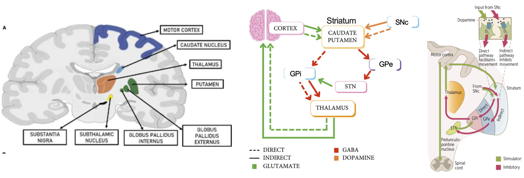 (SNc = substantia nigra pars compacta; SNr = subtantia nigra pars reticulata; GPe = globus pallidus externus; GPi = globus pallidus internus; STN, subthalamic nucleus)
Direct Pathway: The direct pathway begins in the cortex, where excitatory signals are sent to the striatum (caudate and putamen). These structures then send inhibitory signals to the globus pallidus internus (GPi), which inhibits the thalamus. Since the thalamus is responsible for movement, inhibiting an inhibitor results in overall stimulation of movement. Dopamine from the substantia nigra acts on this pathway via D1 receptors, further promoting movement.
Indirect Pathway: The indirect pathway also starts with excitatory signals from the cortex to the caudate and putamen. In response, the caudate and putamen send inhibitory signals to the globus pallidus externus (GPe), which normally inhibits the subthalamic nucleus (STN). When the GPe is inhibited, the STN becomes more active and stimulates the globus pallidus internus (GPi). This leads to the GPi inhibiting the thalamus, resulting in suppression of movement. Dopamine from the substantia nigra influences this pathway through D2 receptors, reducing its inhibitory effects on movement.
Dopamine receptor D1- and D2-expressing medium spiny neurons (D1-MSNs and D2-MSNs) make up 90% of the neurons in the striatum. Both types receive dopamine signals from the SNc, but they have opposite effects on movement. D1-MSNs, which connect to the GPi and the SNr, are part of the direct pathway and help facilitate movement. On the other hand, D2-MSNs, which project to the GPe, are involved in the indirect pathway and work to inhibit movement.
Note: The SNr and GPi are both key output nuclei in the basal ganglia, but the GPi is often highlighted for body (limbs and trunk) movement control, while the SNr focuses more on eye movements and head posture, which is why the SNr is sometimes left out of certain movement models.